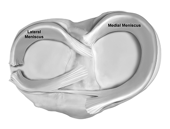
There are many different types of meniscal tears. Common types of traumatic tears include bucket handle tears, flap tears, and radial/root tears.
Broadly, tears can be described as partial thickness, meaning they only affect one side, or full thickness, meaning the tear goes all the way through the meniscus. Tears can be further described based on their appearance. Tears are described as “complex tears” when they have more than one tear pattern. Typically, these involve both a radial and horizontal tear. Finally, the location of the tear has a significant impact on the ability of the tear to heal and the corresponding treatment.
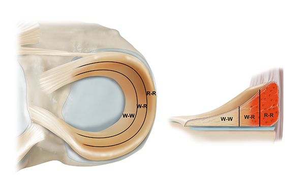
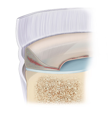
The meniscus has three zones of vascularity. The Red zone is the outer perimeter and has an adequate blood supply to facilitate healing. The Red-White zone is the transitional area in the middle of the meniscus with an intermediate blood supply and capacity for healing. Finally, the White zone is the innermost part with no blood supply and no ability to heal. Tears in the white zone are generally treated by removal of the meniscus, also called a meniscectomy. Tears in the red zone have a good blood supply which provides the necessary biological substances for healing following surgical repair. Tears in the red-white zone need to be assessed to determine whether surgical repair is required for healing.
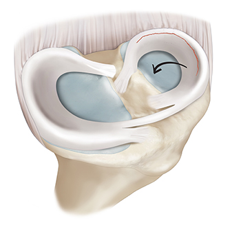
Radial tears are tears that originate from the more central region of the meniscus and extend outwards towards the peripheral edges. Because of their orientation, radial tears disrupt the circumferential protein fibers that allow the meniscus to absorb forces. This can potentially compromise the function of the meniscus. For that reason, surgery is usually required. This pattern of tears is most commonly located in the posterior portion of the medial meniscus or in the middle and anterior sections of the lateral meniscus.
Radial tears can be subdivided into complete tears and incomplete tears. As the tear extends outwards it has the potential to extend all the way to the peripheral rim of the meniscus. If it is able to tear completely across the meniscus and reach this rim it is called a complete radial tear. If it does not reach the rim, it is termed an incomplete radial tear.
Root tears – The meniscal roots are its insertions into the tibia. Each meniscus has an anterior root attachment at the front of the tibia near the kneecap and a posterior root attachment that is near the back of the tibia.
Approximately 10-20% of meniscus tears are root tears. Root tears are a special type of radial tear that occurs within 1 cm of the meniscus attachment or a bony avulsion of the root attachment itself. Root tears lead to extrusion of the meniscus, or the shifting of the meniscus from its natural anatomic position, causing the meniscus to become nonfunctional. Studies have shown that meniscal root tears are biomechanically equivalent to having no meniscus at all, which can accelerate cartilage damage and lead to early progression of osteoarthritis.
Meniscus root tears tend to be traumatic injuries that occur in two groups of patients. The first group is typically younger athletes in their 20s. The second group is adults in their 50s. For both groups, the rapid onset of arthritis associated with these injuries means that surgical repair is typically recommended.
Bucket handle tears are larger tears that occur along the long axis of the meniscus. They run from anterior to posterior along the meniscus, creating a long strip of torn tissue that is separated from the rest of the meniscus, typically displacing into the joint and resembling the handle of a bucket. Bucket handle tears can be acutely debilitating as they are associated with the catching/locking symptoms that trap the knee and prevent patients from straightening out their leg. Like many of the acutely symptomatic tears, bucket handle tears are typically caused by trauma, usually a sporting injury, that occurs when the patient is twisting or pivoting their leg while trying to abruptly change direction. Bucket handle tears are most commonly seen in young athletes, under age 35.
Horizontal tears occur parallel to the meniscus surface. These types of tears can frequently be surgically repaired if they occur in an area of the meniscus that has good blood supply to facilitate healing after the repair. These tears are typically associated with degenerative changes within the meniscus as opposed to acute trauma.
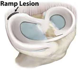
Meniscal ramp lesions are lesions that occur with an ACL injury and lead to damage of the meniscus in the periphery of its posterior segment. This would be the back inner corner of the medial aspect of the knee. These tears will disrupt the attachment between the posterior medial meniscus and the surrounding knee joint capsule.
Ramp lesions have been colloquially termed the “hidden lesion” because they were historically under-recognized. This is in part due to the fact that the tear is located in a portion of the knee termed the “blind-spot.” After an ACL rupture, the knee will have some level of instability in multiple directions. In this situation, the meniscus plays an even larger role in maintaining stability within the knee. Moreover, studies have shown that an ACL reconstruction will be biomechanically inferior to a normal knee if a significant ramp lesion is present but not repaired. While this might be true in a laboratory setting, it is unclear if this remains true in a real-patient context.
Meniscus tears vary in severity and location, influencing treatment decisions. Common types include radial tears, bucket-handle tears, flap tears, and degenerative tears. Each type requires a tailored approach to ensure proper healing and prevent further damage. Dr. Jorge Chahla, leading orthopedic knee surgeon, specializes in diagnosing and treating all types of meniscus tears, helping patients restore knee function with the most effective treatment plans. If you have been diagnosed with a meniscus tear, contact Dr. Chahla’s office in Chicago, Naperville, or Oak Brook to discuss your next steps.