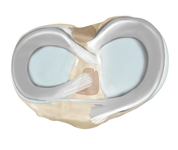
There are three bones that make up the knee joint – the femur (thigh bone), the tibia (shin bone), and the patella (kneecap). The menisci are two C-shaped structures in each knee that are located between the tibia and femur within the knee joint. The menisci play a crucial role in protecting the knee joint by maintaining its stability and cushioning. They are shock absorbers for the knee that are critical to the long-term health and function of the joint.
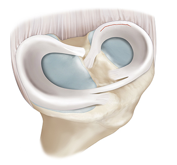
The meniscus is a C-shaped shock absorber located between the tibia and femur within the knee joint. There are two menisci in each knee joint: the medial meniscus located on the inner side, and the lateral meniscus, positioned on the outer side of the knee. When viewed as a cross-section, they are wedge shaped. Their wedge shape allows them to create a perfect fit between the round end of the femur and the flat edge of the tibia within the joint. This fit allows for an even distribution of forces and minimizes regions of high contact pressure. In doing so, the menisci serve a vital role in preventing excessive wear and tear on the articular cartilage. Additionally, the menisci help provide rotational stability to the joint. The menisci have many important roles in the knee, but their ability to absorb forces between the thigh bone and shin bone is the most crucial.
The menisci have a unique composition of protein fibers that allow them to absorb forces. They have parallel fibers that expand circumferentially. They also have fibers that intersect these circumferential fibers at a 90-degree angle. Together, this creates a mesh-like honeycomb network. When you walk, the femur presses down on the tibia. The menisci are between the two and will get compacted, or squeezed, whenever weight is applied to the leg. When the menisci are being compressed, a portion of the force is pushed outwards away from the knee joint due to the unique protein fiber configuration of the menisci. By reducing the amount of force that is transmitted from the femur to the tibia, the menisci will protect the cartilage in the knee. Over years and decades, this cumulative protective effect is crucial, as it will delay or prevent the onset of severe symptomatic osteoarthritis.
Meniscus tears can be acute, meaning they happen suddenly from a single injury, or degenerative, meaning they happen gradually over time with general wear and tear.
Acute meniscus tears are frequently the result of traumatic injuries and can occur during high-risk sports, including soccer, tennis, pickleball, football, or basketball. Typically, injury mechanisms involve aggressive movements in which the knee twists or rotates during pivoting, kneeling, or squatting. Tears can also result from hyperflexion when the knee is flexed beyond its normal range of motion (excessive bending). Everyday activities including getting in and out of a car, using stairs, squatting, or heavy lifting, can also cause tears.
Degenerative tears occur from chronic wear and tear of the meniscus cartilage that accumulates with age. As patients age, the cartilage can become stiff and brittle, leading to degenerative tearing or fraying of the menisci.
Acute meniscus tears are very common knee injuries that frequently affect active individuals. It is not uncommon to suffer a meniscus tear in the setting of a ligamentous injury. In contrast, degenerative meniscus tears are more common in older individuals, affecting 6 out of 10 patients over age 65.
Common symptoms of a meniscus injury include a popping sensation, localized pain and swelling on the inside or outside of the knee depending on the location of the tear. Other symptoms include stiffness, clicking, catching, or locking of the knee. Patients can also report instability, reduced range of motion, and difficulty walking.
Sometimes after 4-6 weeks from the injury, the knee will recover, and symptoms will improve. However, knee pain may return during daily activities including walking, running, and climbing stairs. This recurrent pain may also be associated with symptoms of intermittent swelling, instability, and locking of the knee. Over time, a torn meniscus can predispose the affected knee to the development of osteoarthritis.
The combination of a detailed history, comprehensive physical examination, x-rays, and an MRI (magnetic resonance imaging) is the key to a successful diagnosis of a meniscus injury. Frequently, a torn meniscus can be diagnosed with a physical exam and special tests. Two common specialty exam maneuvers are the McMurray Test and the Apley’s compression test. These maneuvers test for meniscal injury by eliciting pain upon a twisting/rotating motion of the knee. X-rays cannot detect a tear but will identify fractures and degree of arthritis of the knee. An MRI can be ordered to confirm a diagnosis, as well as provide an assessment of the extent of the damage and any associated arthritic or concomitant ligamentous conditions.
A meniscus tear can be treated either non-operatively or with surgery. The tear type, size, and location as well as your age, health, and activity level are all considered when deciding the best treatment for you.
Location plays a key role in deciding the appropriate treatment for a meniscus tear. The meniscus has three zones – the white zone, the red-white zone, and the red zone. The inner portion of the meniscus (white zone) has poor blood supply, which limits the body’s natural healing abilities. Tears in the white zone are generally treated by removing the torn portion (meniscectomy). 30% of the meniscus – the outer edges – has a good blood supply which is essential for the natural healing and repair process. Tears in the red zone may heal with conservative management or can be surgically repaired with confidence that these repairs will heal well.
The meniscus is a crucial structure in the knee that helps absorb shock and provide stability. Meniscus injuries are common and can occur due to sports-related trauma, sudden twisting motions, or gradual wear and tear. Symptoms often include knee pain, swelling, and difficulty moving the joint. Dr. Jorge Chahla, leading orthopedic knee surgeon, offers advanced treatment options to help patients recover from meniscus injuries and regain full mobility. If you are experiencing knee discomfort, schedule an appointment with Dr. Chahla in Chicago, Naperville, or Oak Brook to discuss your treatment options.