FAI FAQs
The femur (thigh bone) fits into the acetabulum (hip bone) to form the femoroacetabular joint or the hip joint. The acetabulum is lined with a thin piece of cartilage that is important for normal hip function (the labrum). The labrum seals the joint, allows for a wider range of movement, and helps keep the bones aligned.
Femoral acetabular impingement, or FAI, is hip impingement caused by a structural problem, specifically bony abnormalities that lead to abnormal contact between the acetabulum and the femoral head. The hip is a ball-and-socket joint comprised of the socket, which is the part of the hip bone called the acetabulum, and the ball, which is the head of the femur. Articular cartilage covers both the ball and the socket and functions to reduce friction, allowing for smooth joint movement. The labrum is a ring of very strong fibrocartilage that functions to tighten the seal between the bones for joint stability; in other words, it acts as a suction cup or rubber band to hold the ball of the femoral head in the socket.
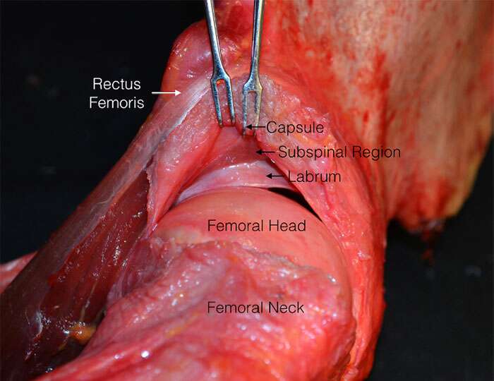
FAI is impingement or pinching of the labrum by the bones in the hip joint, and is a common cause of hip and groin pain in active patients. It can be caused by a developmental deformity or acquired by repetitive movements. The location of the impingement can be described as either the CAM type, Pincer type, or both. CAM type is caused by the abnormal shape of the femoral head and neck, while the pincer type is caused by an abnormal shape of the acetabulum. Due to the anatomical location of the labrum, impingement most commonly leads to injury of the labrum.
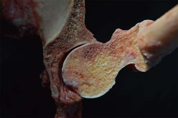
Before Removal of Excess Bone
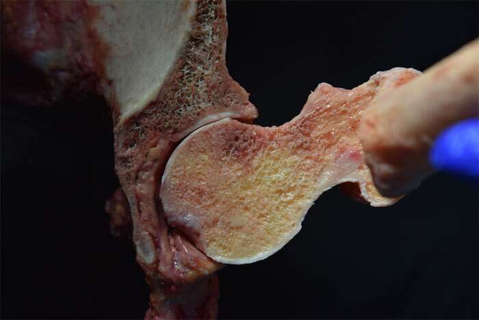
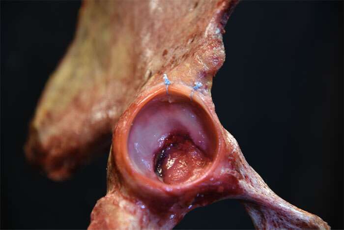
After Removal of Excess Bone
Some people can live active lives, never knowing they have FAI because they do not have any symptoms. However, when FAI symptoms develop, it is usually indicative of damage to the hip cartilage or labrum. Common symptoms of FAI with an associated labral tear include intermittent deep groin pain that is described as an aching pain, pain at the outside of the joint, sharp stabbing pain when twisting, turning, or squatting (as when getting in or out of a car or chair), a dull ache from prolonged sitting or walking, a clicking or locking sound when the hip is moved, instability and decreased range of motion, stiffness and limping. Due to compensation and altered gait mechanics as a result of the symptoms caused by FAI, patients can experience pain in other areas of the body including low back pain, pain in the buttocks, pain in the front of the thigh, and sometimes even knee pain
Yes. FAI pain can be felt in the front of the thigh or down the buttocks. It can also cause low back pain, and sharp pain when getting into or out of a car or a chair.
FAI can be caused by a developmental deformity or acquired by repetitive movements. Some people have a misshapen hip joint that may not cause problems until it is overused and is pushed beyond the hip’s normal range of activity. It is often misdiagnosed as hip bursitis, hip muscle strain, injury due to trauma to the hip joint, or hip dislocations.
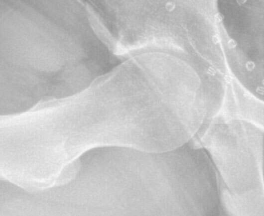
FAI is a clinical diagnosis that incorporates both the symptoms of hip pain that the patient is experiencing as well as the physical exam and diagnostic imaging findings obtained in the sports medicine clinic. During the initial exam in his Chicago sports medicine clinic, Dr. Jorge Chahla or his PA will assess range of motion of the hip joint, strength of the surrounding musculature of the hip, and perform impingement-specific exam maneuvers sensitive for FAI pathology. Additionally, X-rays will be obtained of the hip in several different radiographic views. X-rays can reveal information about the presence of hip dysplasia and arthritis, as well as allow for specific measurements to be taken to assess the radius of curvature of the femoral head and coverage of the femoral head by the acetabulum (hip socket). An MRI may also be warranted which provides information about inflammation, labral tears, and fluid collection. An MRI may be especially useful for determining next steps of care and providing patients with a complete picture of their diagnosis. Lastly, there may be some instances where a CT scan with 3D reconstruction of the hip may be required to look at the morphology of the bony anatomy of the ball and socket joint at a greater level of detail than X-rays can provide.
In terms of differentiating causes of hip pathology, Dr. Chahla and his PA will spend a significant amount of time in his Chicago sports medicine clinic understanding the location and quality of your hip pain and the types of activities that aggravate the hip. The history of the pain, physical exam, and imaging can help differentiate FAI-related pain symptoms from other causes of hip pain including arthritis, gluteal tears, sacroiliitis, psoas tendinitis, adductor tears, and trochanteric bursitis.
Dr. Chahla will employ the hip impingement test during your examination. He will ask you to bring your knee to your chest and then rotate it in toward the opposite shoulder (FADIR [flexion adduction and internal rotation]). If this causes hip pain, it is positive for FAI. However, imaging studies are necessary to confirm the diagnosis. A comprehensive physical exam is key to determine the cause of your pain.
In general, conservative therapy is the preferred initial step in the treatment of FAI. The reason athletes are often diagnosed with FAI is because they overuse the joint in extreme ranges of motion, which damages the labrum and increases inflammation within the joint, therefore, causing pain. Conservative treatment aims to reduce inflammation, minimize the duration and number of acute hip pain episodes, and improve the overall biomechanics of the joint to prevent future hip pain episodes.
In mild to moderate cases, most patients will experience symptomatic improvement with nonsurgical treatment. This involves a change in activities to avoid movements that cause hip pain, using anti-inflammatory medications and ice to reduce hip pain and inflammation, and completing physical therapy. Physical therapy is a key component in treating hip pain. Physical therapy will focus on conditioning, strengthening, posture, and overall improvement in biomechanics, targeting several areas that can help mitigate and often times solve hip pain. Under the guidance of a skilled physical therapist, at Midwest Orthopedics at Rush, who specializes in hip pathology, physical therapy can help strengthen muscles around the hip joint, increase flexibility and maintain range of motion of the joint, decrease the inflammation of the joint or tendons surrounding the hip, and correct hip joint or low back positions that might be adding to your overall pain.
Other modalities of non-operative treatment include in-office injections into the hip joint under ultrasound guidance. Injection options include Cortisone, Hyaluronic Acid (HA), and Hyaluronic Acid (HA) plus Platelet Rich Plasma (PRP). Although injections will not result in correction of the FAI or fix the labral tear, injections work as potent anti-inflammatories that have the potential to decrease the pain symptoms and improve function, allowing patients to get back to their desired activities. Cortisone injections work quickly but will not last as long as PRP and HA injections. Additionally, it is not recommended to have too many repetitive cortisone injections. HA and PRP injections, on the other hand, require about 4-6 weeks on average for the relief to take effect. However, the effects can last up to 7-9 months, and the injections can be repeated. To learn more about injections, contact Dr. Jorge Chahla, Chicago sports medicine surgeon. click here.
Some people live active lives, never knowing they have FAI, and don’t have any problems. By the time FAI symptoms develop there is usually some damage to the hip cartilage (labrum) and with repeated use, the damage will progress. The reason athletes are often diagnosed with FAI is because they overuse the joint in extreme ranges of motion, which damages the labrum more quickly and causes pain.
In mild to moderate cases, FAI symptoms can improve with nonsurgical treatment. This involves a change in activities to avoid those activities that cause pain, including taking time off from athletics, using over-the-counter anti-inflammatory medications to reduce pain and inflammation, and physical therapy. FAI symptoms should resolve within several weeks. Corticosteroid injections can also help relieve hip pain. Chiropractic treatments can sometimes aggravate the condition.
When nonsurgical treatments do not relieve pain and imaging reveals severe tears, detachment of the labrum, or damage to the articular cartilage, FAI surgery will be recommended. FAI surgery can be performed in a minimally invasive procedure called hip arthroscopy, which is usually an outpatient procedure. However, in some cases open surgery may be required. FAI hip surgery is customized for each patient because each patient’s bones and condition are different.
The general recovery process from hip arthroscopy is around 6-9 months after surgery. This is crucial not only to allow for healing of the labrum after repair but also allows patients to slowly progress through a very specific protocol of strengthening, flexibility, range of motion, and stability of the hip. In the immediate post operative period, patients will use crutches and a brace for 2-3 weeks following surgery. Additionally, it is recommended that patients use a continuous passive movement (CPM) machine as well to assist with regaining range of motion in a neutral and safe position. Formal physical therapy will begin on postoperative day 1 or 2 and continue about 2-3 times per week for around 4 months with the expectation that therapy exercises will also be performed by the patient at home in order to allow for maximal functional improvement. Return to sport activities will generally be allowed at 6-9 months after surgery depending on the specific activities that the patient wishes to return to as well as the patient’s overall recovery progress.
Hip preservation refers to the use of procedures to protect and maintain the labrum (the cartilage that lines the hip bone to deepen the hip socket) to prolong the natural lifespan of the hip, to prevent arthritis of the joint, and to avoid or delay hip replacement surgery. FAI predisposes patients to premature hip joint degeneration. Hip replacement surgery is the treatment for advanced arthritis of the hip. Chicago sports medicine surgeon Dr. Jorge Chahla is an expert in hip preservation procedures.
At a Glance
Dr. Jorge Chahla
- Triple fellowship-trained sports medicine surgeon
- Performs over 500 surgeries per year
- Assistant professor of orthopedic surgery at Rush University
- Learn more

