Review of Meniscus Anatomy and Biomechanics
Meniscal tears can cause profound functional, biomechanical, and kinematic derangements within the knee joint, leading to accelerated degeneration of the articular cartilage. The purpose of this review is to summarize the relevant anatomy, biomechanics, and kinematics of the meniscus to guide surgeons during meniscal repair procedures.
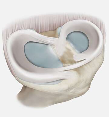
The medial meniscus measures approximately 45.7mm in length and 27.4mm in width, while the lateral meniscus measures approximately 35.7mm in length and 29.3 mm in width. The blood supply to the menisci is provided by branches of the medial inferior, lateral inferior, and middle geniculate arteries. The peripheral regions of each meniscus are the most well-perfused, while the central regions least so. The menisci are attached to the tibia by their anterior and posterior roots, which also function to preserve the menisci’s ability to convert axial (top-down) loads to hoop stress (in-to-out), decreasing the load placed on articular cartilage. The menisci are stabilized by the medial collateral ligament, the transverse ligament, the meniscotibial ligaments, and the meniscofemoral ligaments, and additionally serve as secondary stabilizers of the knee themselves. Additionally, the meniscus plays an important role in normalizing the knee adduction moment (KAM), which correlates with knee medial compartment loading during weight-bearing activities.